Advanced Brain Imaging for Georgia TBI Cases: What Victims Must Know

TBI Advanced Imaging in Georgia: If you or a loved one suffered a head injury in a car accident, truck crash, or other traumatic event in Georgia, you may be facing one of the most frustrating realities in personal injury law: your brain scan came back “normal” — yet you are anything but fine.
Persistent headaches, memory loss, cognitive fog, mood changes, and fatigue are disrupting your life every single day.
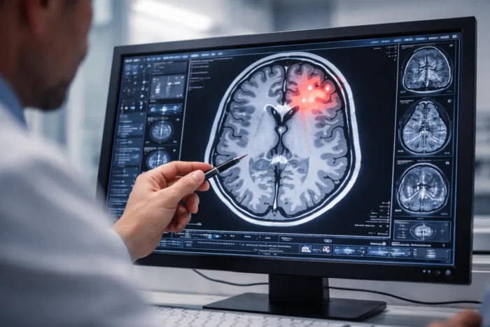
Standard CT scans and conventional MRI can miss the most common type of brain injury entirely. That is why the experienced attorneys at Haug Barron Law Group, Personal Injury Lawyers, serving clients in Decatur, Sandy Springs, and throughout Georgia, work with leading neuroimaging experts to obtain the advanced diagnostic evidence your case demands — and your recovery deserves.
Why Conventional Brain Imaging Falls Short
When emergency room physicians evaluate a head injury, the first tool they reach for is a CT scan. Fast, widely available, and effective at detecting life-threatening bleeds and fractures, CT is the right first step. Standard MRI provides more detail on soft tissue. But for the most prevalent type of traumatic brain injury — mild TBI (mTBI), sometimes called a concussion — both imaging techniques are notoriously unreliable.
The Problem With ‘Normal’ Scans
The brain’s gray matter — the neuron cell bodies — often remains structurally intact after a traumatic event. What gets damaged is the brain’s white matter: the axonal fiber tracts that carry electrical signals between regions. This type of injury is called diffuse axonal injury (DAI). Conventional CT and MRI images do not visualize white matter connectivity. They produce, in essence, a photograph of the brain’s architecture — not a map of its wiring.
The result? Insurance companies and defense attorneys routinely point to a “negative” scan as proof that there is no serious injury. Meanwhile, their clients’ victims are struggling to hold down jobs, maintain relationships, and live normal lives.
According to research published in the American Journal of Neuroradiology and endorsed by the American College of Radiology, advanced neuroimaging techniques are specifically indicated for patients with mild TBI whose conventional CT and MRI findings are negative but who continue to experience persistent symptoms — exactly the patients most at risk of being disbelieved and undercompensated.
Advanced Brain Imaging Technologies Explained
Over the past decade, neuroscience has developed a suite of imaging modalities that go far beyond the static picture of a conventional MRI. Each technology reveals a different dimension of brain function and connectivity. In the right legal hands, they can transform an “invisible” brain injury into compelling, objective courtroom evidence.
Diffusion Tensor Imaging (DTI)
DTI is arguably the most powerful and widely litigated advanced imaging tool available today. Rather than photographing brain structure, DTI measures the movement of water molecules along axonal fiber tracts — the highways of the brain’s communication network. In a healthy brain, water diffuses in a highly organized, directional pattern. After traumatic injury, disrupted or severed axons cause water to diffuse in a scattered, disordered way that DTI captures as measurable abnormality.
Key brain regions commonly affected in TBI — including the corpus callosum, frontal lobes, and internal capsule — are invisible to standard MRI but clearly visualized on DTI. A DTI scan that reveals disrupted white matter tracts in areas consistent with the mechanism of injury (e.g., a rear-end collision) can be powerful corroborating evidence of your injury.
Why it matters for your case: DTI findings provide objective, measurable data — fractional anisotropy (FA) values — that a qualified neuroradiologist can present in a deposition or at trial. It takes the injury out of the realm of subjective complaint and into the realm of documented pathology.
Functional MRI (fMRI)
While DTI maps the brain’s structural wiring, fMRI maps the brain’s activity. By tracking blood oxygen levels (the BOLD signal) in different brain regions, fMRI can detect areas that are underactive, overactive, or failing to communicate properly with other regions — all common sequelae of traumatic brain injury.
Two fMRI approaches are used in TBI evaluation: task-based fMRI (measuring how the brain responds to cognitive tasks like working memory) and resting-state fMRI (measuring baseline connectivity across brain networks, including the default mode network). Disruption of the default mode network, for example, has been consistently associated with cognitive symptoms in mTBI patients.
Quantitative EEG (qEEG)
Quantitative EEG analyzes the brain’s electrical activity patterns and compares them against normative databases to identify statistically abnormal brain wave patterns. For TBI cases, qEEG can reveal connectivity disruptions and changes in cognitive processing speed that correlate directly with the patient’s reported symptoms. As a functional measure, it complements the structural data from DTI.
PET and SPECT Scanning
Positron emission tomography (PET) and single photon emission computed tomography (SPECT) measure metabolic activity and blood flow in the brain, respectively. Regions with reduced glucose metabolism or blood perfusion — even when structurally intact — can demonstrate physiological dysfunction consistent with TBI. These modalities are particularly useful in cases involving chronic post-concussive syndrome or repeated head trauma.
MR Spectroscopy (MRS)
MRS measures the concentrations of specific neurochemicals — including N-acetylaspartate (NAA), a marker of neuronal health — within brain tissue. Reduced NAA levels in the absence of visible structural damage on conventional MRI can indicate neuronal injury or dysfunction consistent with mTBI.
Advanced Imaging and Georgia Personal Injury Litigation
Understanding the science is only half the battle. To win your case, advanced imaging evidence must be strategically obtained, properly preserved, and effectively presented to a judge and jury. This is where having an experienced Georgia personal injury attorney becomes essential.
Admissibility Under Georgia and Federal Standards
Georgia courts, like federal courts operating under Daubert standards, require that expert scientific testimony be grounded in reliable methodology. Advanced neuroimaging evidence — particularly DTI — has been the subject of significant litigation over admissibility. Courts across the country have generally found DTI evidence admissible when presented by a qualified neuroradiologist or neurologist who can explain the methodology, its acceptance in the scientific community, and its application to the specific patient.
The key is the quality of the expert and the quality of the imaging protocol. Not all DTI scans are created equal — acquisition parameters, field strength, and post-processing methodology matter enormously. At Haug Barron Law Group, we work with imaging facilities and medical experts who follow rigorous, peer-reviewed protocols that hold up under cross-examination.
Defense Tactics You Should Expect
Insurance defense attorneys are well-versed in attacking advanced neuroimaging evidence. Common tactics include:
- Challenging the qualifications of the imaging expert
- Arguing that the findings reflect pre-existing conditions rather than the accident
- Pointing to white matter changes as non-specific or age-related
- Citing the “insufficient evidence for routine clinical use” caveat in medical literature — without disclosing that this refers to population-level research, not individual clinical findings
- Hiring their own neuroimaging experts to reinterpret scans favorably
Anticipating and effectively countering these strategies requires experience with TBI litigation specifically — not just general personal injury practice. Our attorneys have the depth of knowledge to respond to these challenges and protect the integrity of your medical evidence.
Timing Matters: Why You Must Act Quickly
Advanced imaging, when performed promptly after an injury, captures acute changes in white matter integrity. Some DTI abnormalities are most detectable in the days and weeks following injury; others persist and evolve over months. Delay in obtaining advanced imaging — whether due to lack of awareness, inadequate early medical care, or delayed legal representation — can mean the loss of critical evidence.
If you have suffered a head injury in an accident, do not assume your “normal” CT scan is the end of the story. Contact our office as soon as possible so we can evaluate whether advanced neuroimaging is warranted in your case.
TBI in Truck Accident Cases: A Particular Challenge
Traumatic brain injury is especially common — and especially severe — in Georgia truck accident cases. The physics of a collision involving an 80,000-pound commercial vehicle are orders of magnitude more violent than a typical passenger car crash. The forces involved in truck accidents — sudden deceleration, rotation of the head, secondary impacts — are precisely the mechanisms most associated with diffuse axonal injury.
Yet truck accident TBI cases are also some of the most heavily contested by insurance carriers and trucking companies. With potentially catastrophic damages at stake, defense teams spare no expense in challenging causation, disputing the severity of injury, and attributing symptoms to pre-existing conditions. This makes the objective evidence provided by advanced imaging not just helpful, but often essential.
At Haug Barron Law Group, Personal Injury Lawyers, we handle complex truck accident litigation throughout Georgia. Our team understands the interplay between federal motor carrier regulations, trucking company liability, and the medical evidence required to prove the full extent of your injuries — including brain injuries that standard scans miss.
What to Do If You Suspect a TBI After a Georgia Accident
If you were involved in a serious accident and are experiencing any of the following symptoms — even if your initial CT or MRI was reported as “normal” — you may have a traumatic brain injury that requires advanced evaluation:
- Persistent headaches that do not resolve
- Memory problems or difficulty concentrating
- Sensitivity to light or noise
- Sleep disturbances
- Mood changes, irritability, or depression
- Dizziness, balance problems, or visual disturbances
- Fatigue disproportionate to the demands of your day
- Slowed processing speed or “brain fog”
These are the hallmarks of mild TBI — and they can be documented with the right imaging. Here is what you should do:
Step 1: Seek Immediate Medical Attention
See your primary care physician, a neurologist, or a concussion specialist and report all symptoms in detail. Ask about referral for advanced neuroimaging if conventional scans have not explained your symptoms.
Step 2: Document Everything
Keep a daily symptom journal. Note your cognitive performance at work, social engagement, sleep quality, and any limitations on activities you could previously perform easily. This chronological record is valuable evidence.
Step 3: Contact an Experienced Georgia TBI Attorney
Do not accept a low settlement offer from an insurance company before your injury is fully evaluated and documented. Once you release your claims, you cannot reopen them — even if your symptoms worsen. The attorneys at Haug Barron Law Group offer free consultations and represent TBI victims on a contingency fee basis, meaning you pay nothing unless we recover for you.
Still Have Questions About Traumatic Brain Injury Claims in Georgia?
A “normal” CT or MRI result does not mean your brain injury isn’t real — and it should not prevent you from pursuing full and fair compensation. If you are wondering how advanced imaging is used as legal evidence, what your rights are after a TBI, or how Georgia law applies to your situation, our Frequently Asked Questions page provides clear, straightforward answers to help you understand your options.
Additional Resources
The following peer-reviewed and professional resources informed the preparation of this article and may be helpful to individuals researching traumatic brain injury:
- American Journal of Neuroradiology — Imaging Evidence and Recommendations for TBI: Advanced Neuro- and Neurovascular Imaging Techniques
- Journal of the American Academy of Psychiatry and the Law — Advanced Neuroimaging and Mild TBI Litigation, Revisited
- Centers for Disease Control and Prevention — Traumatic Brain Injury & Concussion
A “normal” brain scan does not mean your injury isn’t real — and it shouldn’t mean you walk away without the compensation you deserve. At Haug Barron Law Group, our attorneys work with leading neuroimaging experts to uncover the evidence that standard scans miss and build a compelling case that reflects the true extent of your traumatic brain injury. Contact us today for a free consultation and don’t let an insurance company use an incomplete diagnosis to undervalue your claim.
Legal Disclaimer
This article is provided for informational purposes only and does not constitute legal advice. Reading this article does not create an attorney-client relationship with Haug Barron Law Group. The results of any prior case do not guarantee or predict the outcome of any future matter. If you have been injured, please consult with a qualified attorney regarding the specific facts of your situation.
Contact Haug Barron Law Group Today for a FREE Consultation.